Applicability of Proton Therapy Treatments
Proton Therapy can make a difference in a large variety of cancers.
Because of the physical and biological properties of Protons, all tumors which have been traditionally treated with X-rays can be irradiated with protons. These are therefore all tumors that were previously using linear accelerators (with and without intensity modulated radiotherapy – IMRT) or related equipment with X-rays (this includes Rapid Arc and CyberKnife).
Video -link: Conditions treated by Proton Therapy
- Children
Children have priority in the indications for proton therapy because the frequency of radiation-induced second cancers can be reduced. Radiation damage to growing organs, such as growth plates and other functional organs can be avoided or reduced to the extent that their functions are retained (especially in the brain, eyes, ears and skull base region). For more information on the treatment of children, see ..
Proton beam therapy of pediatric patients

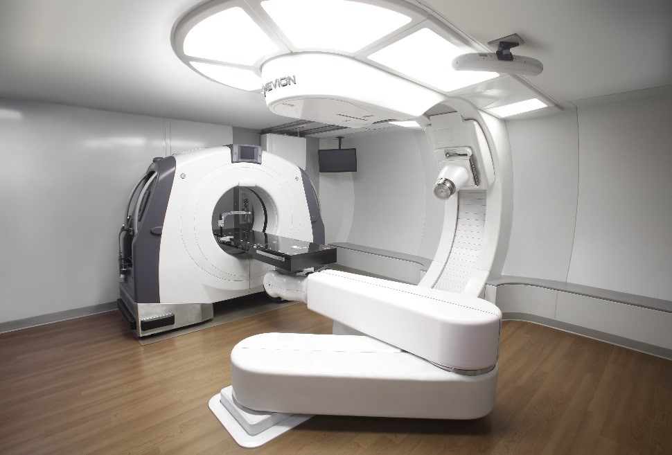
Source: ProCure Training and Development Center
Tumors that are particularly suitable for Proton Therapy are tumors that are hardly treatable due to the high side effects with X-rays.
- Tumors of the head and neck
- Brain and skull base tumors

Proton beam therapy for brain tumour compared to other radiation techniques.
Source: Tarbell, Yock, Loeffler MGH-FBPTC, HMS

Proton beam therapy for skull base tumour compared to other radiation techniques.
Source: ProCure Training and Development Center.
- Tumors of the eye
- Lung and liver tumors

Proton beam therapy of non small cell lung cancer compared to other radiation techniques.
Source: ProCure Training and Development Center.

Adapted from Chang JY, et al. Int J Radiat Oncol Biol Phys. 2006:65(4):1087-1096.
Gy=standard measure of absorbed radiation; CGE=cobalt gray equivalent.
- Tumours of abdomen and pelvis
- Prostate cancer

Proton beam therapy for prostate cancer compared to other radiation techniques.
Source: ProCure Training and Development Center
Compared with IMRT/X-rays, proton therapy delivers:
- 35% less radiation to the bladder
- 59% less radiation to the rectum
 Adapted from Vargas C, et al. Int J Radiat Oncol Biol Phys. 2008;70(3):744-751.
Gy=Gray, the standard measure of absorbed radiation; CGE=Cobalt Gray Equivalent

- Tumors and metastases in the spine

Proton beam therapy for tumor next to spine compared to other radiation techniques.
Source: ProCure Training and Development Center.
- Local recurrence and individual metastases
- Rectal and Anal Cancers
Clinical data is limited regarding the efficacy of proton irradiation in rectal and anal cancers. For more than 30 years, the standard of care for this cancer has been radiation therapy to the primary site and draining the lymphatics delivered concurrently with aggressive chemotherapy. While this is a successful regimen, the risk of significant toxicity is high.
A small comparative treatment-planning study of proton and X-ray radiation therapy indicated that proton therapy, used either as the sole modality or as a boost to X-rays, has potential advantages over conventional X-rays used alone in the treatment of inoperable patients with a large rectal cancer. The proton therapy treatment plan is more effective in sparing the small bowel, the bladder, and the femoral heads. Higher doses are possible with proton therapy and may allow for downstaging and potentially curative resection in patients with locally advanced disease. ( Isaacson U, Montelius A, Jung B, and Glimelius B. Comparative treatment planning between proton and X-ray therapy in locally advanced rectal cancer. Radiother Oncol. 1996;41(3):263-272. ; Meyer JJ, Czito BG, Willett CG. Particle radiation therapy for gastrointestinal malignancies. Gastrointest Cancer Res. 2007;1(suppl 2):S50-59.)

- Not suitable tumors are
For the time being, postoperative breast irradiation is still not suitable.
There is hope that with better accuracy and protection of the surrounding tissue proton radiation will be able to reduce the side effects and late sequelae such as the occurrence of lung cancer (lung cancer) or a higher incidence of coronary atherosclerosis and myocardial infarction in the left breast irradiation. Theoretically, this is possible due to the better accuracy of irradiation with protons. For this to work, not only the patient but also the breast itself must be positioned exactly the same for each exposure. Such a precise bearing is so far very difficult due to the high mobility of not only the breast but also the skin of the entire chest area. We are currently working on a storage concept that corrects the irradiation using lasers for surface profile comparisons to store three-dimensional shapes to ensure that each individual exposure sessions remains sufficiently identical to the original location, in order to enable even breast cancer patients a gentle irradiation of![]() the breast.
the breast.